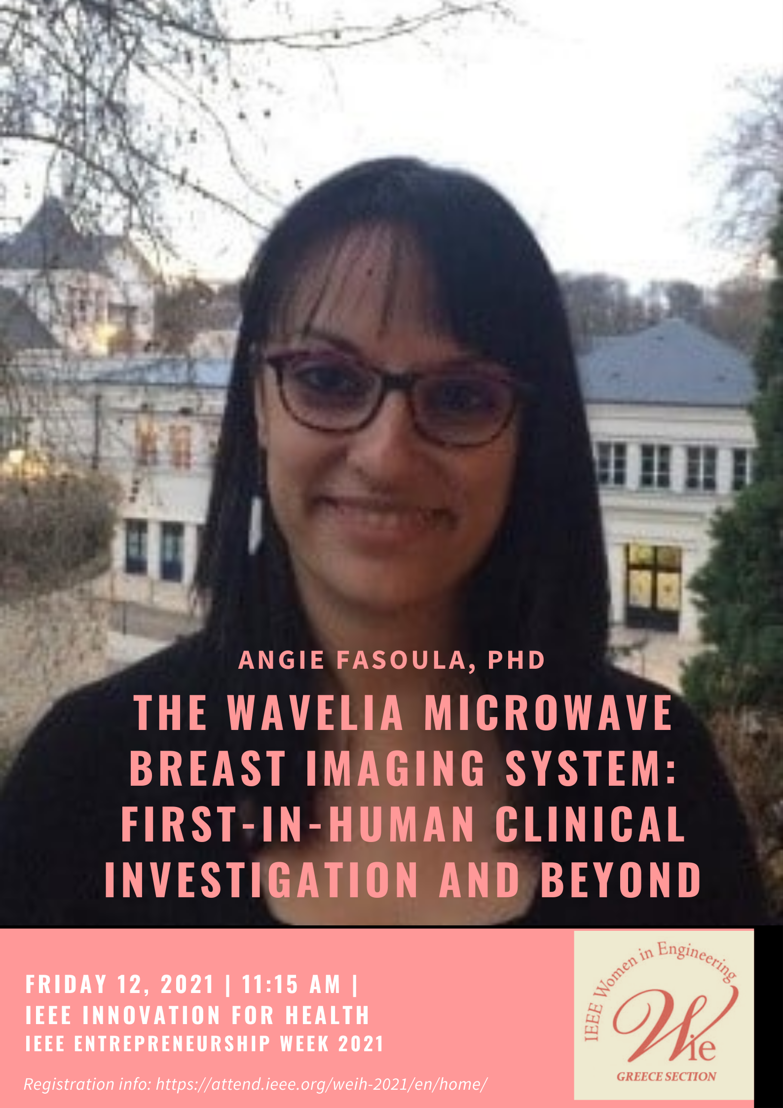
Coffee Talk with Dr Angie Fasoula

Dr. Angie Fasoula is the Technical and Scientific Manager of the Wavelia Microwave Breast Imaging system development www.wavelia.com , in MVG Industries. She holds a PhD degree in Electrical and Computer Engineering from TUDelft, The Netherlands (2011) and an Electrical and Computer Engineering Diploma from the Aristotle University of Thessaloniki, Greece (2005). In 2006, she was awarded a 3-year Marie Curie fellowship for early-stage research, hosted in Thales Nederland BV, in Delft, where she partially carried-out her PhD research. Her thesis was focused on radar signal processing and imaging algorithm development for target classification. Since late 2013 she is dedicated to R&D work on Microwave Breast Imaging, within MVG Industries. Prior to that, she performed post-doctoral research in brain network connectivity analysis, using MagnetoEncephaloGraphic (MEG) data, in the Brain and Spine Institute, Paris, France (2011-2013).
She also gained 2-year industrial experience as a Radar System Engineer, in Thales Air Systems, Limours, France (2009-2011).
The Wavelia Microwave Breast Imaging system: First-In-Human Clinical Investigation and beyond
Microwave Breast Imaging (MBI) is an emerging technology that has potential to detect diseased breast tissue, and thus offer a non-invasive, non-ionizing and lower-risk adjunct for breast cancer diagnosis. MBI is based on the contrast in dielectric properties between cancerous breast tissue (high water content) and healthy breast tissue (low water content) in the microwave frequency spectrum.
WaveliaTM is an MBI system prototype of 1st generation, which demonstrated the ability to detect dielectric contrast between tumour phantoms and synthetic fibroglandular tissue in preclinical studies and has recently completed a First-In-Human (FiH) clinical investigation on a 25-symptomatic patient cohort, hosted in NUIG Clinical Research Facility Galway, Ireland. In this study (ClinicalTrials.gov NCT03475992), the Wavelia MBI system was evaluated in the clinical setting for the first time, using mammography as the reference conventional imaging modality and post-surgery histology data to assess the size of the cancers. Ultrasound, MRI and core biopsy data was also collected as reference, where available as part of the patient’s standard-of-care. In this clinical setting, Wavelia showed preliminary promise to detect and approximate underlying breast abnormalities to the appropriate location, in patients with palpable biopsy-confirmed invasive carcinomas and benign breast lesions, such as cysts and fibroadenomas. The device also demonstrated promising results in delineating the size and malignancy risk of the detected breast lesions.
The FiH study results of Wavelia will be presented during this talk, organized in 3 axes: (a) Breast lesion detectability, (b) Breast lesion sizing, (c) Discrimination between malignant and benign breast lesions. The methodology that was employed in the Wavelia semi-automated Quantitative Imaging Function (QIF) during this FiH study, to support morphological breast lesion detection based on persistence, lesion sizing and lesion characterization in a low-dimensional feature space, spanning shape and texture-based features, will be also outlined and the critical design parameters highlighted.
The promising findings from the FiH study provided initial data to support the valid clinical association of the technology and warranted the preparation of further clinical investigations, with an upgraded prototype version of the Wavelia system (Wavelia #2) to progressively address the identified technological challenges. Larger and more diverse patient datasets are needed to validate these findings and delineate the cases where the Wavelia MBI modality may offer a beneficial adjunct to current diagnostic protocols. The technological status and the current vision on the clinical potential of Wavelia will be briefly discussed, before closing this talk.

